

The Next Generation of PRP: From Quantity to Functional Precision
Platelet-Rich Plasma (PRP) has become a widely utilized tool in Regenerative Medicine, with platelet concentration and total platelet dose well established as essential parameters for achieving therapeutic benefit. These metrics have provided a strong foundation for standardizing PRP and ensuring the delivery of sufficient bioactive mediators to support tissue repair. At PLYMOUTH MEDICAL, we have long been proponents of characterizing cellular dosing in PRP and we were the first to introduce point-of-care cell counters to help clinicians quality control their PRP in 2017.
However, variability in clinical outcomes suggests an opportunity to further refine how PRP is characterized and optimized. Emerging evidence indicates that PRP preparations with similar platelet counts can demonstrate significant differences in biological activity, influenced not only by patient-specific factors but also by how the product is processed, handled, and activated. [1]
These insights prompt an important evolution in our understanding of PRP: while platelet dose remains a necessary foundation, optimizing the functional quality and coordinated signaling of these platelets may further enhance therapeutic outcomes.
Expanding the Framework: From Platelet Count to Functional Biology
Platelets themselves are not the therapeutic agent, they function as delivery vehicles.
Within their alpha granules, platelets store and release a complex network of growth factors, cytokines, and signaling molecules that orchestrate tissue repair. These include platelet-derived growth factor (PDGF), transforming growth factor beta (TGF-β), vascular endothelial growth factor (VEGF), and insulin-like growth factor (IGF), among others.
As emphasized in recent reviews, the regenerative capacity of PRP is influenced not only by platelet number, but by the molecular content and functional integrity of these bioactive mediators. [1]
Importantly, these signaling molecules operate within tightly regulated biological systems where:
Relative ratios matter, as imbalances can shift signaling toward fibrosis rather than regeneration
Timing matters, as release kinetics influence cellular recruitment, angiogenesis, and matrix remodeling
More is not always better, as excessive concentrations may lead to receptor saturation or disorganized signaling
The clinical efficacy of PRP is therefore shaped not only by platelet number, but by:
The quality of platelets
The integrity of their bioactive cargo
The timing and coordination of growth factor release
Platelet count remains a useful and practical proxy for PRP potency, but growth factor profile, activation method, leukocyte content, and patient-specific factors ultimately determine the true biological and clinical effect as a whole.
New Orthobiologics Preparations: From the lab to the clinic
Standard PRP
Conventional PRP preparation methods are primarily designed to concentrate platelets from whole blood. While approaches may vary, the process typically ends once the platelet-rich fraction is isolated.
This model emphasizes platelet yield and concentration, which, while important, does not seem to fully account for the functional quality or biological coordination of the final product as suggested in recent papers. As a result, variability in platelet integrity, activation state, and growth factor availability may exist despite similar platelet counts.
Platelet-Rich Plasma Concentrate
Platelet-Rich Plasma Concentrate represents a natural evolution of PRP preparation, building upon established principles of platelet concentration and dose while incorporating additional processing steps in sterile medical devices to further refine the biological composition and functional quality of the final product prior to reinjection.
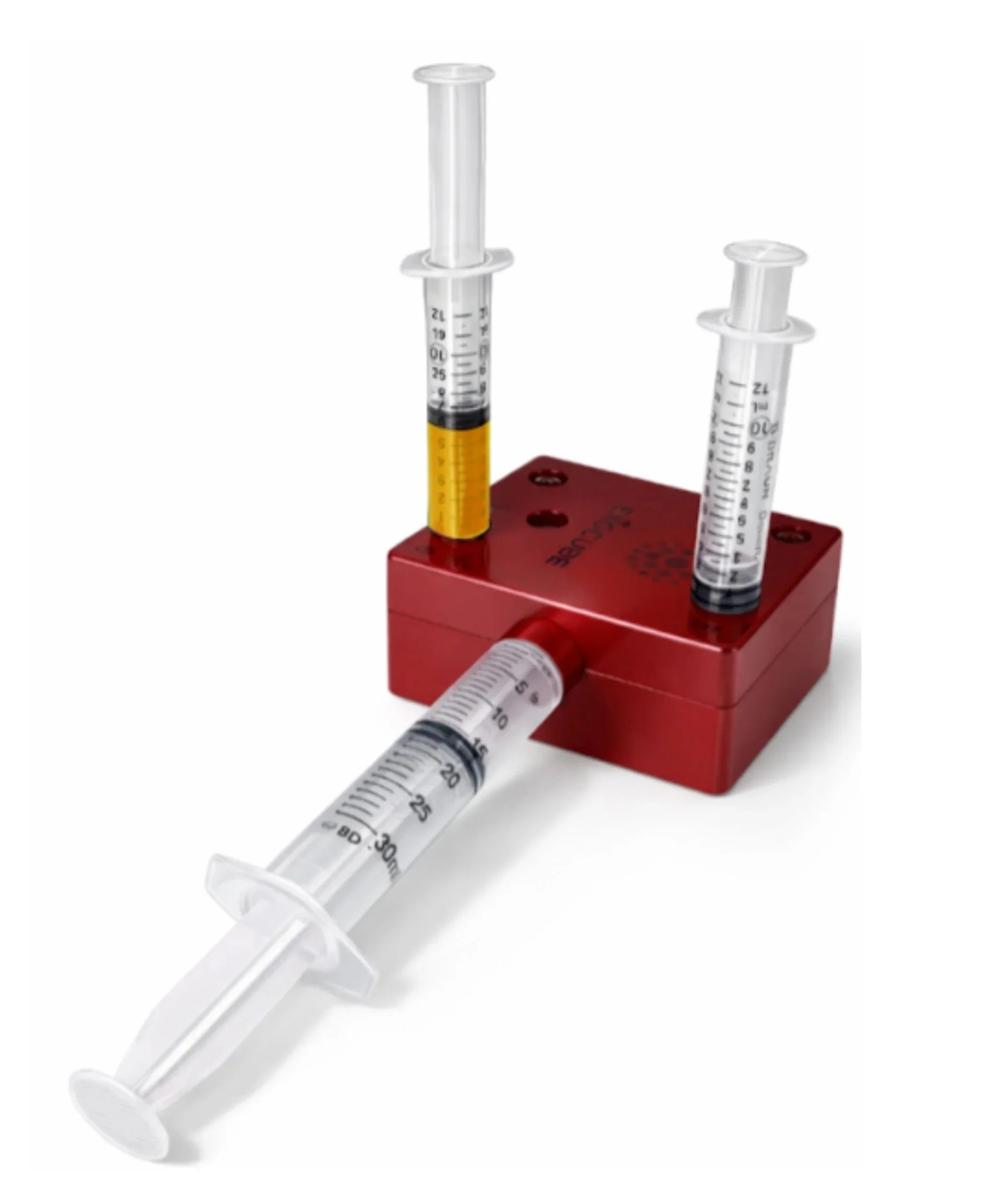
A controlled, multi-step approach that includes activation, filtration, and concentration, is now available with devices such as the Exocube™ to further refine the final biological product into a platelet-rich injectate with growth factors, cytokines and concentrated plasma proteins.
1. ACTIVATION
Following PRP preparation, platelet activation becomes a critical step in defining how growth factors are released.
Activation can be achieved through biochemical approaches such as thrombin or calcium chloride, thermal approaches involving freeze-thaw cycles known to create platelet lysates, as well as surface-mediated mechanisms including collagen-based systems and glass bead activation. Each method influences the timing, magnitude, and efficacy of growth factor release.
Mechanical, surface-mediated activation approaches such as using glass beads utilize a negatively charged glass surface to trigger platelet activation through contact with the intrinsic pathway, a mechanism that has been well described in foundational platelet biology literature. [2] Glass beads promote platelet adhesion and aggregation by mimicking the surface characteristics of damaged tissue or foreign material, initiating a physiologic activation response. [3] This leads to platelet degranulation and the release of growth factors such as PDGF, TGF-β, and VEGF.
Mechanical activation of monocytes using medical-grade glass beads is a well-characterized technique in autologous cytokine therapy, most notably employed in systems such as the Orthokine/Autologous Conditioned Serum (ACS) method developed by Dr. Peter Wehling. In this process, whole blood is incubated in syringes containing borosilicate glass beads, which provide a contact surface that mechanically stimulates monocytes and other leukocytes through integrin-mediated adhesion signaling. This contact activation triggers an anti-inflammatory and regenerative cytokine cascade. Studies have demonstrated significant upregulation of interleukin-1 receptor antagonist (IL-1Ra), the primary therapeutic cytokine of interest, which competitively inhibits the pro-inflammatory effects of IL-1β at the receptor level. [5-7] Additionally, elevated levels of interleukin-10 (IL-10), an anti-inflammatory cytokine, and various growth factors including hepatocyte growth factor (HGF), insulin-like growth factor-1 (IGF-1), platelet-derived growth factor (PDGF), transforming growth factor-beta (TGF-β), and fibroblast growth factor (FGF) have been reported in the conditioned serum. [7–9] Notably, while some pro-inflammatory cytokines such as IL-1β and tumor necrosis factor-alpha (TNF-α) may also increase during incubation, the ratio of anti-inflammatory to pro-inflammatory mediators — particularly the IL-1Ra to IL-1β ratio — is heavily skewed toward an anti-inflammatory milieu, which is believed to underpin the therapeutic benefit observed in conditions such as osteoarthritis and tendinopathy. [6,8,10]
Beyond immediate activation, surface contact-based systems contribute to clot formation, which may serve as a biologic scaffold that modulates the spatial and temporal release of signaling molecules.
2. FILTRATION
Filtration introduces an additional layer of refinement by removing unwanted cellular debris and optimizing the plasma fraction.
Multi-step filtration processes are designed to:
Improve the consistency of the final product
Reduce, though not completely, the particulate matter that may interfere with signaling
Reduce pro-inflammatory cellular components and improve the overall biologic profile
Enhance clarity and injectability of the final preparation
In systems that utilize surface-mediated activation, such as glass bead platforms, filtration may also help manage residual particulate material to ensure a cleaner final plasma fraction following processing. Though not 100% effective at removing all cells and platelets, some filters can be used to selectively remove debris.
While not intended as a sterilization method, filtration contributes to a more controlled biological environment by refining both the physical and cellular composition of the final product. [1]
3. PLASMA CONCENTRATION (DEHYDRATION)
Polysulfone (PSf) hollow fiber membranes have emerged as a critical enabling technology in the preparation of next-generation orthobiologic formulations, particularly for the concentration of plasma proteins from platelet-poor plasma (PPP) by dehydration. In the Orthobiologics context, a new generation of biocompatible ultrafiltration devices employing hollow fiber semipermeable membranes have been developed to concentrate PPP proteins by reducing plasma water in a controlled manner, retaining molecules with a molecular weight larger than the membrane pore size along with several important extra-platelet growth factors, producing a low-volume, viscous, protein-rich plasma product. [14] Specifically, these devices exploit the fact that insulin-like growth factor-1 (IGF-1) and hepatocyte growth factor (HGF) are mainly present in the PPP fraction outside of platelets, and newly developed ultrafiltration technologies can concentrate plasma proteins such as fibrinogen, alpha-2-macroglobulin (A2M), IGF-1, HGF, cytokines, and other biomolecules. [14] The recent Mayo Clinic study by Showel, Sellon, Boettcher et al. validated this approach, demonstrating that ultrafiltration of PPP is a reliable, quantitative method for concentrating biologically active A2M, achieving mean A2M concentrations of 6.66 mg/mL in A2M-rich plasma (A2MRP) versus 1.56 mg/mL in baseline plasma, with IGF-1 similarly concentrated to 257.63 ng/mL from a baseline of 68.82 ng/mL. [13] The molecular weight cut-off (MWCO) of the polysulfone membranes is a key design parameter: A2M, at approximately 720 kDa, is readily retained alongside other large therapeutic proteins, while smaller pro-inflammatory cytokines and plasma water pass through. [15]
This selective concentration mechanism allows clinicians to harvest a therapeutically enriched product from what was previously a discarded PRP byproduct, creating a complementary anti-catabolic therapy that can be used alongside or independently of PRP to address the protease-driven degradation central to osteoarthritis pathophysiology. Although there is a paucity of research on plasma based protein enrichment via polysulfone filters, such as for alpha-2 macroglobulin–rich plasma (A2M), the market is rife with filter products. We encourage our readers to ensure the devices they are using are properly classed and approved by the FDA.
Through plasma filtration with polysulfone filters, one can:
Increase the concentration of bioactive molecules, including growth factors and plasma-derived signaling mediators
Enrich and concentrate regulatory proteins such as α2-macroglobulin (A2M), a broad-spectrum protease inhibitor that has been shown to attenuate cartilage-degrading enzymatic activity and slow the progression of post-traumatic osteoarthritis, highlighting the dual regenerative and protective roles of plasma-derived bioactive fractions. [4]
Preserve biological activity during processing
Maintain the integrity of key signaling components
Emerging literature in cartilage and joint preservation biology further supports the concept that plasma-derived preparations represent complex, multi-component biologic systems rather than single-factor therapeutics. These systems include not only growth factors but also regulatory proteins and enzymatic modulators that collectively influence extracellular matrix turnover, inflammation, and cartilage homeostasis. [13] This evolving understanding shifts the paradigm from simple concentration of platelets or plasma volume toward intentional modulation of the biologic environment, optimizing both regenerative signaling and tissue-protective mechanisms.
Closing: From Quantity to Molecular Precision
Platelet concentration and total platelet dose have long served as foundational parameters in PRP therapy, providing an essential framework for standardization and clinical use. Building on this foundation, growing evidence suggests that biological outcomes are also influenced by the functional quality of platelets and the signaling environment they create.
Growth factors, cytokines, and other bioactive mediators stored within platelets remain central to tissue repair, with their activity influenced by both intrinsic platelet characteristics and external processing variables.
Evidence indicates that platelet function may vary with factors such as age, metabolic status, and oxidative stress, while processing methods can further shape the preservation and expression of bioactive signals. These do not diminish the importance of platelet concentration, but rather highlight additional considerations contributing to variability in clinical response.
The future of PRP lies in a more integrated biological framework, one that considers platelet dose alongside growth factor composition, platelet integrity, patient-specific factors, and the processing methods used to prepare and refine the final product. This approach supports a more nuanced understanding of PRP as a biologic system, with multiple interdependent variables contributing to therapeutic effect.
Ultimately, the evolution of Orthobiologics will not be defined solely by platelet quantity, but by how effectively biological signals are preserved, structured, and delivered to support healing.
-The PLYMOUTH MEDICAL Education Team
Citations
04/21/2026